MBST is the only therapeutic medical device that uses the same physical principle as the MRI known from diagnostics: magnetic resonance. Sounds good, but what does that actually mean?
Simply put, MRI and MBST have a similar technological basis. The way in which both devices work is based on the fact that they contain components that can generate magnetic resonance conditions with the tissue in the body. But they differ in what they want to achieve with the use of magnetic resonance. One has a diagnostic aim, the other a therapeutic one.
The aim of MRI
Put simply, a magnetic resonance tomograph is like a very large digital camera that can take photos of the inside of the body. These images show the different tissues of the body in such detail that doctors can see whether there is an inflammatory reaction in a “photographed” area for example, or whether it is healthy or diseased tissue, such as tumour tissue. To generate these images, hydrogen protons, a strong magnetic field, radio wave pulses and very powerful computers are required. One advantage of this type of image generation is that the patient is not exposed to radiation during the examination, as is the case with X-rays or CT scans, for example.
How does an MRI work?
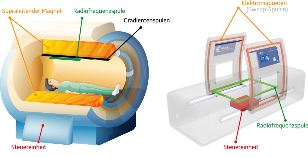
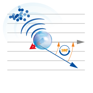
An MRI device (also known as MRI tomograph or MRI scanner) is a very large device in which the patient lies. In its usually ring-shaped tunnel, often referred to as a tube, a very strong magnetic field is generated in order to influence the hydrogen protons in the patient’s body. When the magnetic field is switched on, the hydrogen protons in the patient’s body align themselves parallel to the magnetic field. The MRI device then emits precisely timed radio wave pulses, which stimulate the hydrogen protons by transferring energy in such a way that they change measurably. This means that the hydrogen protons are practically nudged energetically and as a result change their position. They tilt once around their axis by 180° during which they absorb energy. When the impulse is switched off, the hydrogen protons rotate back to their original position. In doing so, they release some of the previously absorbed energy back into the surrounding tissue.
Diagnostic use of magnetic resonance technology
The amount of energy which is emitted by the hydrogen protons can be measured. A computer uses this data to generate images from underneath the skin. The time required for the hydrogen protons to turn back is known as relaxation time. As the tissues in the body have different water content (bones less than cartilage for example), the tissues also contain different amounts of hydrogen protons. Due to the differing relaxation times of the measured tissue types, MRI technology can visualize the differences in the form of image contrasts. From the large amount of data of the measured signals, the computer generates an image in different shades of gray using special mathematical procedures. By changing the measuring settings, certain types of tissue can be enhanced or attenuated in the resulting image, depending on what shall be examined.
Therapeutic use of magnetic resonance technology
In MBST magnetic resonance therapy, the same thing happens at the start: a magnetic field is generated, the protons align themselves with the field lines and they absorb and release energy radio wave pulses are switched on and off again.
Continue reading how the developer of MBST therapy came to the conclusion that this process could also have a therapeutic effect: History of MRI
Years of interdisciplinary research and development work were necessary before magnetic resonance could also be used therapeutically. The proton densities and relaxation times of tissues were determined with magnetic resonance spectroscopy. For example, tissue-specific relaxation times were measured in close cooperation with the Institute of Nuclear Physics at the University of Giessen. Tissue-specific cell parameters were determined together with the Laboratory for Medical and Molecular Biology at Aachen University of Applied Sciences. From this basis, the sequences for the tissue-specific MBST therapy cards were developed. The aim is to trigger various processes to achieve an effect on cell metabolism. Scientific data indicates that MBST magnetic resonance technology stimulates various biophysical processes and could trigger anti-inflammatory and pain-relieving effects.
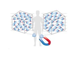
Why do both MRI and MBST use hydrogen protons?
Hydrogen is the most common element in the human body and at the same time the most sensitive component for magnetic resonance. By using hydrogen protons, the largest possible amount of protons contained in the tissue can be addressed.
Differences between the MBST magnetic resonance therapy system and magnetic resonance imaging (MRI)
How can you get a good MRI image and how does that influence MBST therapy?
- MRT
An MRI image of a foot for example shows the details of the tissue: the ankle, the bones of the lower leg, the muscles in the calf or the skin. This detailed image is achieved by using different MRI sequences and the different weighting to bring tissue-specific characteristics/differences into prominence for diagnostic purposes.
- MBST
MBST therapy uses different pulse sequences to address a specific tissue. MBST magnetic resonance technology transfers energy to the tissue that shall be treated, which is intended to activate endogenous processes.
Magnetic field strengths, gradients and the resulting noise
- MRT
A magnetic resonance scanner is very large and requires a specially shielded room. No radio waves or electrical devices shall interfere with the examination which would have a negative effect on image quality.
MRI does not expose the patient to a uniform strong magnetic field but it is stronger on one side of the body and weaker on the other. This results in a magnetic field gradient, i.e. only a thin layer of protons is addressed. If this layer is subjected to a radio pulse of the corresponding Larmor frequency, only these protons are activated and only their signals are registered by the receiving coil.
MRI technology generates further gradients across and at right angles to the recording plane. These are subdivided into columns and rows and can be read out like a table. Each time these gradient fields are switched on and off, the so-called “Lorentz forces” act on the suspensions of the coils, causing the knocking noises typical of MRI.
- MBST
The magnetic strengths and electrical energies required for MBST therapy do not need to be nearly as strong as for MRI. Since MBST does not want to generate images, it does not need the loud gradient coils for spatial mapping. The magnetic resonance field used by the MBST therapy device is approx. 10,000 times weaker than the fields used in an MRI. MBST magnetic resonance technology uses fast adiabatic passage technique, with which magnetic resonance can be generated with weak magnetic fields. This is a method for reversing nuclear spin orientations and a prerequisite for nuclear magnetic resonance.
“The generation of repeated spin resonance sequences according to the principle of fast adiabatic passage allows the generation of nuclear magnetic resonance conditions in weak magnetic fields. This means that the nuclear magnetic resonance condition can also be used for small and medium-sized systems. The method is used in nuclear magnetic resonance therapy. Three interacting magnetic fields are required for nuclear magnetic resonance therapy: firstly a static main magnetic field, secondly a modulated magnetic field parallel to this and thirdly an alternating field that satisfies the Larmor condition and is perpendicular to the other two. The magnetic field strength of the modulated magnetic field then passes around the static field while the frequency remains constant. As the strength decreases, the alternating field is also activated. The aim is to correlate the frequency of the modulated magnetic field with the spin-lattice relaxation time. The typical magnetic field is generated in a Helmholtz coil.” (https://de.wikipedia.org/wiki/Schnelle_adiabatische_Passage)
The advantage for patients is that MBST therapy devices work quietly, virtually silently, because far fewer magnets are needed.
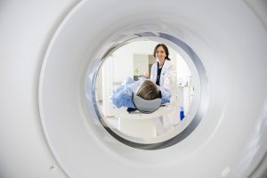
Claustrophobia and the need to lie still
- MRT
During an MRI scan, patients usually lie in a narrow tube and have to remain as still as possible for the duration of the examination. This is because movement can blur the resulting images, so that the images would have to be repeated. This would mean that the patient would have to remain in the MRI scanner for an even longer time. Patients with claustrophobia or other forms of anxiety are therefore sometimes lightly sedated during the examination.
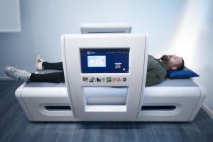
- MBST
The MBST therapy devices have an open design. There is no narrow tunnel, only some applicators located next to the body (the exact construction depends on the respective therapy device). Even in the whole-body therapy device, the lying surface is not narrow or enclosed. As the aim are not sharp detailed images, it is not necessary to lie without moving. The patient can relax on the lying surface during the treatment and, for example, read, listen to music or even sleep.